Single-Stage Wound Repair Using Human Reticular ADM
Case series of 5 trauma patients shows single-stage reconstruction with reticular acellular dermal matrix achieves durable closure in 2.5 months.
Damon Ebanks
Medipyxis
Medical education note: This article is for clinicians and is not a substitute for patient-specific medical advice.
Quick Take
A retrospective 5-patient case series evaluated single-stage wound reconstruction using human reticular acellular dermal matrix (ADM) (with selective use of a placental membrane allograft) across complex trauma wounds of the ankle, foot, forehead, and scalp. Mean wound size was 37 cm² (30–49 cm²). Wounds spanned clean-contaminated (class 2) to dirty/infected (class 4). The average time to closure was 2.5 months, no postoperative complications were reported, no unplanned returns to the OR occurred, and all patients achieved satisfactory closure at one year—even over previously exposed bone.
Why it matters: For trauma and oncologic defects where flap options are limited—or where you want to avoid staged coverage—reticular ADM can enable single-stage closure with durable outcomes when paired with meticulous debridement and wound bed prep.
Study at a Glance
- Design: Retrospective case series, 5 adults (Mar 2024–Jan 2025). Primary outcomes: time to closure, complications, and unplanned reoperation.
- Etiology & sites: All injuries were motor-vehicle–related; sites included ankle, foot, forehead, scalp. Mean wound size 37 cm².
- Contamination class: Included class 2, 3, and 4 wounds.
- Grafts used: Human reticular ADM and, in select cases, a placental membrane allograft (SomaGen® Meshed; Salera® Mini Membrane, MTF Biologics).
- Outcomes: 2.5-month mean closure, no postoperative complications, no unplanned OR, durable closure at 1-year follow-up, including areas with prior bone exposure.
Limitations: Small sample, retrospective design, mixed graft types; results are hypothesis-generating.
Why Human Reticular ADM for Single-Stage Repair?
Reticular ADM (HR-ADM) preserves the architecture of the reticular dermis, yielding a porous, vascular-friendly scaffold that supports cell infiltration, neovascularization, and rapid re-epithelialization—properties documented in preclinical work where HR-ADM accelerated closure while minimizing excess granulation.
Broader systematic and narrative reviews underscore that acellular dermal matrices can be reliable adjuncts for complex wounds across the upper and lower extremities, especially when conventional options (local flaps, free tissue transfer) are constrained by patient status or defect geometry.
Who's a Good Candidate?
- Trauma defects with exposed tendon/bone after thorough debridement and infection control.
- Mixed-contamination fields (class 2–4) when staged flaps are impractical and the team can ensure aggressive bed prep and surveillance.
- Cosmetically sensitive sites (forehead/scalp) where single-stage contour preservation is preferred.
- Medically complex patients where minimizing anesthesia and procedures is strategic.
Single-Stage Protocol
Goal: Immediate biologic coverage that integrates and epithelializes without returning to the OR for staged grafting.
1) Debridement & Bed Readiness
Debride to a bleeding, clean bed; remove biofilm and devitalized tissue. Culture/treat infection per standard of care.
2) Matrix Selection & Preparation
Choose reticular ADM sized to the defect; meshed/fenestrated options (as used in the series) aid fluid egress and conformability.
3) In-Situ Placement
Ensure full contact with the wound bed; secure edges per your protocol (sutures/staples/adhesive strips). Consider a non-adherent contact layer and moisture-balanced secondary dressings.
4) Adjuncts
Based on exudate and location, use compression for venous disease (if arterial supply permits) and consider NPWT as a bolster in high-shear zones.
5) Follow-Up Cadence
Frequent early checks (first 2–3 weeks) to confirm adherence and vascular ingrowth; then space visits as epithelialization progresses. In the case series, average closure was 2.5 months.
Outcomes to Discuss With Patients and Teams
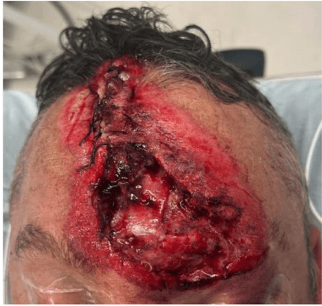
 Patient with motor vehicle collision injury, 11 months post-op — durable closure with HR-ADM.
- Time to closure: ~2.5 months on average for single-stage HR-ADM cases in this series.
- Reoperations/complications: None reported post-op; no unplanned OR.
- Durability: Satisfactory 1-year closure across all cases, including over bone.
- Cost & access: ADMs carry material costs; however, reducing staged procedures may offset total episode costs in selected patients—an area for future prospective research.
How This Aligns With the Broader Evidence
- Mechanism: HR-ADM scaffolds support rapid keratinocyte migration, cellular infiltration, and angiogenesis—observed in a diabetic murine excisional model.
- Clinical scope: Systematic and narrative reviews support ADM use across complex extremity wounds and reconstructive scenarios, citing integration and contour maintenance as advantages when flaps are not feasible.
- Experience in chronic wounds: Evidence syntheses describe HADM as a useful ECM scaffold in chronic, protease-rich environments.
FAQ
What makes "reticular" ADM different?
It preserves the reticular dermis' open architecture, which supports faster epithelial coverage and neovascularization versus denser scaffolds in preclinical models.
Does single-stage ADM mean no skin graft later?
Often yes—that's the goal. In this series, cases closed without unplanned OR and achieved durable closure at one year; however, staged grafting remains appropriate for some wounds.
Can I use HR-ADM in contaminated or infected wounds?
It can be used after adequate debridement and infection control. The series included class 3–4 wounds with favorable outcomes, but careful selection and close follow-up are essential.
Bottom Line
For difficult trauma defects—including those with prior bone exposure—human reticular ADM can enable single-stage closure with no unplanned reoperations in carefully selected cases. Start with aggressive debridement, ensure a clean, vascular bed, choose reticular ADM that conforms and vents fluid, and follow closely during the first weeks as epithelialization takes hold.
References
- Advancing Wound Reconstruction: Single-Stage Repair using Human Reticular Acellular Dermal Matrix (ADM). Case series; n=5, mean 37 cm² wounds, 2.5-month mean closure.
- Dolivo D, Xie P, Hou C, et al. Application of decellularized human reticular allograft dermal matrix promotes rapid re-epithelialization in a diabetic murine excisional wound model. Cytotherapy. 2021.
- Iorio ML, Shuck J, Attinger CE. Wound healing in the upper and lower extremities: a systematic review on ADM use. Plast Reconstr Surg. 2012.
- Tognetti L, Pianigiani E, Ierardi F, et al. Human ADM in advanced wound healing and surgery: state of the art. Dermatol Ther. 2021.
- Kirsner RS, Bohn G, Driver VR, et al. Human acellular dermal wound matrix: evidence and experience. Int Wound J. 2015.