NPWTi-d for Infected Abdominal Dehiscence After Surgery
Learn how NPWTi-d with hypochlorous acid accelerates cleansing, clears infection, and supports closure of large dehisced abdominal wounds after body-contouring surgery.
Damon Ebanks
Medipyxis
Medical education note: This article is for clinicians. Always follow device IFUs, local protocols, and individual patient factors.
Managing Infected, Dehisced Abdominal Wounds After Body-Contouring Surgery: Early Outcomes Using NPWTi-d
Why This Matters
Full-thickness abdominal wound dehiscence after abdominoplasty or liposuction is a serious complication that can lead to prolonged hospital stays, increased risk of incisional hernia, and repeat operations—especially when infection and slough stall progress.[1,2]
Negative pressure wound therapy with instillation and dwell (NPWTi-d) of a topical wound solution has been shown to automatically cleanse wound surfaces, solubilize devitalized tissue, remove infectious exudate, and reduce bacterial load—key goals when managing large, contaminated abdominal wounds.[1,3]
A recent three-case experience describes the use of NPWTi-d with hypochlorous acid (HOCl) via reticulated open-cell foam with through-holes (ROCF-CC) as an adjunctive pathway to rapidly convert infected, dehisced abdominal wounds to clean, granulating beds and then transition patients to outpatient care.[1]
Key Takeaways (At a Glance)
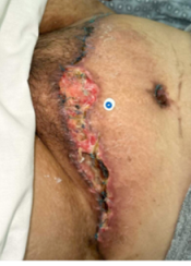 Patient with an infected abdominal dehiscence after body-contouring surgery.
- Population: Three patients with massive, infected, full-thickness abdominal dehiscence after elective body-contouring surgery. Infection was confirmed by non-contact, real-time fluorescence imaging.[1,4]
- Therapy used: NPWTi-d via ROCF-CC foam; HOCl solution; −150 mmHg continuous pressure; instillation every 2.5–3.5 hours; 10–15-minute dwell; dressing changes three times per week.[1,3]
- Starting point: Baseline slough covered 15–50% of the wound surface; post-debridement wound volumes ranged from 130.6 to 1,186.1 cm³.[1]
- Outcomes: Infection clearance and discharge in a mean of 8.7 days (range 7–11). All wounds progressed to closure after transition to conventional NPWT; total healing time 7.9–12.8 weeks.[1,2]
What Is NPWTi-d and Why Use It Here?
NPWT with instillation and dwell intermittently bathes the wound with a topical solution and then resumes negative pressure. During instillation and dwell, the solution loosens slough, fibrin, and biofilm; during suction, the solution and solubilized debris are removed through the foam, supporting hydromechanical debridement while maintaining a moist wound environment.[3,5]
Consensus guidelines and real-world data suggest NPWTi-d with ROCF-type dressings can accelerate wound-bed preparation, reduce signs of local infection, and shorten time to readiness for closure compared with traditional NPWT alone in complex, contaminated wounds.[3,5]
For large, infected, dehisced abdominal wounds, this hydromechanical effect is particularly valuable: each cycle targets nonviable tissue and bioburden without requiring constant operative returns, while negative pressure helps manage edema and exudate.[1,3]
How the Team Ran NPWTi-d (Clinic Playbook)
1. Surgical Debridement + Antibiotics
All patients underwent sharp surgical debridement and received systemic antibiotics before or along with NPWTi-d. Infection was confirmed using non-contact fluorescence imaging at admission.[1,4]
2. Dressing Choice and Settings
A ROCF-CC foam dressing (with through-holes) was used with HOCl instillation at −150 mmHg continuous pressure. The team set cycles to instill every 2.5–3.5 hours with a 10–15-minute dwell, and dressing changes were scheduled three times per week—parameters that fall within published NPWTi-d protocols using HOCl solutions.[1,6]
3. Ongoing Imaging at Each Change
At every dressing change, clinicians used fluorescence imaging to visualize areas of high bacterial load and NIRS to evaluate tissue oxygenation (StO₂) in and around the wound. Fluorescence imaging has been shown to improve detection of clinically significant bacterial burden, while NIRS provides non-invasive perfusion data that can guide escalation of vascular or local therapies.[4,7]
4. Transition Plan
NPWTi-d was discontinued as soon as infection cleared (based on imaging and clinical exam). Patients were then discharged and continued healing under conventional NPWT in the outpatient setting.[1,2]
Results
Across the three cases, infection cleared and patients were medically ready for discharge within 7–11 days (mean 8.7 days), supporting the idea that a focused NPWTi-d phase can substantially shorten the inpatient portion of the course for large, infected abdominal wounds.[1]
Once infection was controlled and NPWTi-d was stopped, all wounds went on to closure within 7.9–12.8 weeks under standard NPWT, mirroring healing trajectories reported in larger NPWTi-d dehiscence series.[1,2]
Clinically, serial images show dense slough and contamination at admission, rapid clearance of devitalized tissue under NPWTi-d, and the emergence of robust granulation tissue before transition to outpatient NPWT—a pattern consistent with other Veraflo Cleanse Choice case collections in complex abdominal wounds.[1,8]
Mechanism in Action: Hydromechanical Debridement
Authors attribute progress in part to hydromechanical removal of nonviable tissue through the ROCF-CC dressing during instillation cycles: each HOCl dwell solubilizes debris, and the subsequent negative-pressure phase evacuates contaminated fluid and slough through the dressing's through-holes.[1,3]
Systematic and consensus reviews of NPWTi-d highlight similar effects—enhanced wound cleansing, reduced bioburden, and faster development of a clean granulating base—especially when complete surgical debridement of a large, complex wound is difficult in one session.[3,5]
Practical Pearls for Dehisced Abdomens
Confirm infection objectively. Non-contact fluorescence imaging helps verify bioburden and track response between changes, reducing reliance on subjective clinical signs alone.[4,9]
Don't skip debridement. Sharp surgical debridement plus systemic antibiotics before or with NPWTi-d remains foundational; NPWTi-d is an adjunctive wound-bed preparation tool, not a replacement for source control.[1,3]
Be consistent with cycles. Maintaining a tight instillation interval (2.5–3.5 hours) and dwell time (10–15 minutes) helps sustain hydromechanical cleansing between nurse visits.[1,5]
Know when to switch. Stop NPWTi-d as soon as infection clears and the wound shows a clean, granulating base; then transition to conventional NPWT or other closure strategies.[1,2]
Who Is a Good Candidate?
Patients with large, infected, surgically dehisced abdominal wounds after body-contouring procedures who can tolerate negative pressure and instillation cycles—and for whom debridement and appropriate antibiotic therapy are already underway—may benefit from a short NPWTi-d phase followed by outpatient NPWT.[1,3]
Selection should always account for device-specific contraindications and NPWTi-d guidelines (e.g., exposed organs or vessels, uncontrolled bleeding, untreated osteomyelitis), along with patient comorbidities and goals of care.[3,5]
Limitations
This is an initial, uncontrolled three-case series; outcomes may not generalize across all abdominal wounds, reconstructive techniques, or patient risk profiles.[1,2]
Commercial involvement and device selection (e.g., specific NPWTi-d system and ROCF-CC dressing) should be considered when interpreting usability and performance claims, as most published NPWTi-d experience is tied to branded platforms.[3,8]
References
-
Desvigne M, Gonzales C. (CS-006) Management of Infected Dehisced Abdominal Wounds with Adjunctive Negative Pressure Wound Therapy and Instillation of a Topical Wound Solution. SAWC Spring 2025 Poster. SAWC
-
Porfidia R, et al. Treatment of wound dehiscence utilizing negative pressure wound therapy with instillation and dwell time (NPWTi-d). Wounds. 2020;32(2):E13–E18. PubMed
-
Silverman RP, et al. Negative Pressure Wound Therapy With Instillation and Dwell Time: Mechanisms and Evidence. Plast Reconstr Surg Glob Open. 2023;11(8):e5180. PMC
-
Armstrong DG, et al. Point-of-care fluorescence imaging reveals extent of bacterial load in diabetic foot ulcers. Int Wound J. 2023;20(2):554–566. PubMed
-
Acosta J, et al. Effectiveness of Negative Pressure Wound Therapy With Instillation and Dwell Time and ROCF-CC: Real-World Evidence. Wound Repair Regen. 2025. PMC
-
Kirkland KB, et al. Utilizing Instilling Negative Pressure Wound Therapy with Hypochlorous Acid–Based Wound Cleanser. Surg Sci. 2023;14(7):259–267. SCIRP
-
Račytė A, et al. Oxygen saturation increase in ischemic wound tissues during therapy monitored by NIRS. Biomedicines. 2024;12(8):1805. PMC
-
3M. V.A.C. Veraflo Therapy Cleanse Choice Dressing Case Study Collection. 2020. Wounds International
-
Price N, et al. Routine fluorescence imaging to detect wound bacteria reduces antibiotic use and antimicrobial dressing expenditure while improving healing rates. Diagnostics. 2020;10(11):927. MDPI
-
McElroy EF, et al. Use of negative pressure wound therapy with instillation and dwell time using a reticulated open-cell foam dressing with through-holes. Int Wound J. 2019;16(5):1188–1194. Wiley