Polycythemia Vera Leg Ulcer: Multimodal Case & Protocol
JAK inhibition and anti-biofilm care reversed an 83.5% area reduction in a chronic PV-associated leg ulcer after hydroxyurea discontinuation.
Damon Ebanks
Medipyxis
Medical education note: This article is for clinicians and is not a substitute for patient-specific medical advice.
Quick Take
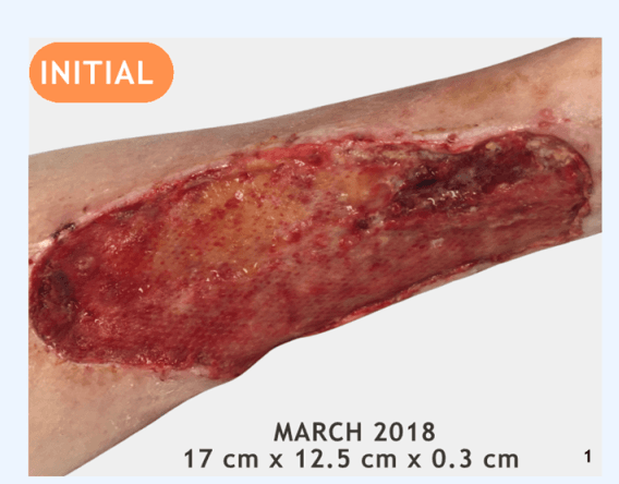
 76-year-old female with chronic non-healing ulcer to lower-left extremity, initial visit.
A 76-year-old woman with polycythemia vera (PV) and a chronic lower-extremity ulcer improved only after two pivotal changes: stopping long-term hydroxyurea and starting ruxolitinib for systemic PV control, and adding cadexomer iodine to aggressively target suspected biofilm in the wound bed.
From an initial wound size of 17 × 12.5 × 0.3 cm in 2018, the ulcer ultimately decreased to 7.6 × 4.6 × 0.1 cm by early 2025—an 83.5% area reduction—after hematologic optimization, de-escalation of hydroxyurea, introduction of ruxolitinib, and sustained anti-biofilm dressing use.
Case at a Glance
PV was diagnosed in 2014, and the leg ulcer began in March 2018 on the lower extremity. The patient also had prolonged exposure to World Trade Center (9/11) dust and toxins.
Before the turning point, multiple standard therapies were tried with only partial or transient benefit: hyperbaric oxygen therapy (HBOT) for ischemia, serial low-frequency ultrasound (LFU) sessions, collagenase and calcium alginate dressings, and a bilayered bioengineered skin substitute.
Biopsies and sharp debridement provoked bleeding and subsequent wound worsening, underscoring how PV-related vascular dysfunction and hydroxyurea-related skin toxicity can translate into leg ulcers that are unusually fragile and slow to granulate.
The major inflection points were discontinuation of hydroxyurea with initiation of ruxolitinib in 2019, followed by escalation of topical anti-biofilm therapy with cadexomer iodine in 2023.
Why PV Ulcers Stall
PV is characterized by erythrocytosis, leukocytosis, and thrombocytosis, leading to increased blood viscosity and microvascular dysfunction. Randomized data from the CYTO-PV trial show that keeping hematocrit below 45% significantly reduces major cardiovascular and thrombotic events.
Hydroxyurea (HU), a cornerstone cytoreductive agent in higher-risk PV, is also linked to a distinctive pattern of painful ankle and lower-leg ulcers that often resolve only after the drug is discontinued.
For patients whose PV is controlled but who develop nonhealing leg ulcers while on HU, case reports describe healing when HU is stopped and the patient is transitioned to ruxolitinib, a JAK1/2 inhibitor used as a second-line option in HU-intolerant or HU-resistant PV.
Local Wound Strategy That Worked
Given the chronicity of the ulcer and its behavior on sharp debridement, the local plan prioritized biofilm control with low-trauma methods. After an initial response to LFU plateaued, cadexomer iodine became the workhorse topical agent.
Because the ulcer bled readily and flared after biopsies and aggressive sharp debridement, subsequent debridement was restrained and individualized, using noncontact LFU, gentle curettage, and autolytic or enzymatic methods to avoid further injury.
Clinician-Ready Protocol: PV-Associated Chronic Leg Ulcer
1) Stabilize the Hematology First
Aim for hematocrit under 45% and ensure guideline-concordant use of low-dose aspirin and phlebotomy.
If an ulcer emerges or stalls on hydroxyurea—especially in the characteristic malleolar distribution—discuss HU interruption or discontinuation and second-line options such as ruxolitinib with hematology.
2) Map Vascular Status
Obtain ABI and/or toe pressures and assess for venous hypertension and arterial disease.
3) Control Biofilm and Moisture
For sloughy, exudative PV ulcers, cadexomer iodine is a practical first-line topical because it combines sustained iodine release with an absorbent starch matrix.
4) Debridement—Go Gently
In PV patients with friable microvasculature and HU-related skin fragility, consider noncontact or low-trauma debridement options first.
5) Reevaluate Every 2–4 Weeks
Track wound area, depth, pain, and granulation; if not trending toward smaller size within 2–4 weeks, escalate both systemic and local care.
Bottom Line
For PV-associated leg ulcers, durable progress typically requires aligning systemic and local strategies: optimize hematocrit and antithrombotic therapy, reconsider HU when ulcers stall, and pair that with deliberate biofilm-focused local care.
References
- CYTO-PV trial. NEJM. 2013.
- Sirieix ME, et al. Leg Ulcers and Hydroxyurea. Arch Dermatol. 1999.
- Hydroxyurea-induced leg ulcers: case reports.
- Cadexomer iodine for chronic wounds: systematic review and meta-analysis.
- Ruxolitinib in HU-intolerant PV.