Nonhealing 'Abscess' After I&D? Think Epidermoid Cyst (and Plan Excision)
Chronic post-drainage wounds may conceal epidermoid cysts. Complete capsule excision, not repeated drainage, offers definitive healing.
Damon Ebanks
Medipyxis
Medical education note: This article is for clinicians and is not a substitute for patient-specific medical advice.
The "Abscess" That Wouldn't Heal
If managing a nonhealing abscess wound persisting weeks after incision and drainage (I&D) despite appropriate cultures, dressings, and debridement, consider expanding the differential diagnosis. Chronic or recurrent "abscess wounds" on the back, shoulders, buttocks, scalp, axillae, or hair-bearing lines often mask an epidermoid (sebaceous) cyst—or a retained cyst wall following prior I&D. Complete excision of the cyst capsule typically provides definitive healing, not additional drainage or antibiotics.
Two Fast Cases from Clinical Practice
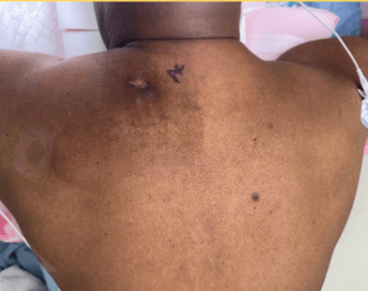
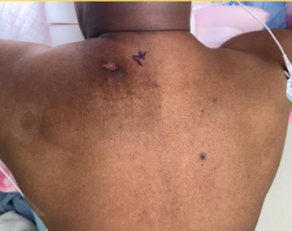 Case 1: Sebaceous cyst, initial visit.
Case 1 – The Slow Burn: A 59-year-old man with smoking history developed a back abscess, underwent I&D, then spent 3–4 months in conservative care with serial debridements and daily dressings. The wound hardened and plateaued. Surgical consultation led to en bloc excision of the sebaceous cyst and nonhealing wound with layered complex closure. Pathology confirmed cyst diagnosis, and he healed without complications.
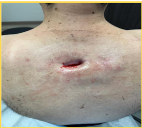 Case 2: Sebaceous cyst, initial visit.
Case 2 – The Boomerang: A 41-year-old man with uncontrolled diabetes and heavy smoking experienced recurrent drainage after I&D, progressive fibrosis, and no forward progress. Surgery removed the wound and underlying cyst; closure used local rotation advancement flap plus incisional negative-pressure wound therapy (iNPWT). He improved despite minor postoperative epidermolysis.
Why These Cases Matter: Both "abscess wounds" were fundamentally cyst problems. The dressing approach or antibiotic regimen was not the limiting factor—the capsule itself was.
What We Tend to Miss
"Sebaceous cyst" is a misnomer. These lesions are typically epidermoid/epidermal inclusion cysts (EICs) loaded with keratin, not sebum. The sac features stratified squamous epithelium lining. When ruptured or incompletely removed, keratin incites a foreign-body granulomatous response mimicking a chronic abscess.
I&D serves as a bridge, not a cure. During acute inflammation, the cyst wall is friable and surgical planes are indistinct; complete removal becomes difficult and recurrence risk rises. Once inflammation subsides, complete excision of the intact capsule represents the definitive step and carries the lowest recurrence risk.
Clues suggesting a cyst or retained wall include:
- Central punctum
- History of "cheesy" keratinous drainage
- Location in hair-bearing zones
- Prior lump that "ruptured"
- Failure to progress despite weeks of thoughtful wound management
Imaging Pearls
Bedside ultrasound helps when physical findings remain ambiguous. Classic epidermoid cyst features include an ovoid, usually heterogeneous or mildly hyperechoic lesion with posterior acoustic enhancement and dermal connection. In ruptured cysts, the "submarine sign"—a tract or projection toward the epidermis—differentiates from simple abscess.
Management That Moves the Needle
Acute Flare (Inflamed or Infected Lesion)
When frank infection and fluctuance are present, I&D is appropriate with antibiotics guided by systemic signs. However, do not rely on I&D as definitive therapy for epidermoid cysts.
The Stalled "Abscess Wound"
After a reasonable window of wound care (2–4 weeks) without trajectory toward closure, escalate to surgical evaluation for complete capsule excision. Timing matters: excise once inflammation subsides.
Surgical Considerations
Goal: Remove the entire cyst lining; include the punctum to minimize recurrence.
Closure strategies: Use layered closure for tension control. In higher-risk wounds (smoking, diabetes, flap or rotation closures), consider prophylactic iNPWT over closed incisions to reduce surgical-site complications.
Decision Aid: "The Abscess That Lingers"
- Identify nonhealing "abscess wound" ≥2–4 weeks after I&D.
- Reexamine for a punctum or prior lump history.
- Use bedside ultrasound if available—look for the submarine sign.
- If suspicion is high, avoid repeat I&D; schedule elective excision once inflammation cools.
- For high-risk closure—consider iNPWT for 5–7 days.
- Reinforce risk-factor control.
Bottom Line
When an "abscess wound" refuses to heal, reframe the question: "What structure is sustaining this?" In many cases, the answer is a cyst wall—and once removed, the wound finally behaves like a wound again.
References
- IDSA SSTI guidelines on incision and drainage.
- Surgical excision of epidermal inclusion cysts.
- Incisional NPWT in high-risk closures.
- Ultrasound features of epidermoid cysts and abscesses.