Hydromechanical Debridement With NPWTi-d for Limb Salvage
Learn how NPWTi-d with hypochlorous acid accelerates debridement, reduces bioburden, and supports limb salvage in complex diabetic lower-extremity wounds.
Damon Ebanks
Medipyxis
Medical education note: This article is for healthcare professionals and not a substitute for device IFUs or clinical judgment.
Overview
Lower-extremity ulcers in patients with diabetes are difficult to heal because of neuropathy, ischemia, infection, and necrotic tissue; they carry a high risk of lower-extremity amputation when not optimally treated. Large reviews estimate that 50–60% of diabetic foot ulcers (DFUs) become infected and that a substantial proportion of these infections progress to major amputation if not controlled.[1,2]
A recent case series by Alonso and colleagues describes the use of Negative Pressure Wound Therapy with Instillation and Dwell (NPWTi-d) using hypochlorous acid (HOCl) instilled through a reticulated open-cell foam dressing with through-holes (ROCF-CC) as an adjunctive limb-salvage strategy for complex diabetic lower-extremity ulcers. In this protocol, NPWTi-d was integrated into a structured diabetic limb-salvage pathway rather than used in isolation.[3,4]
Study Snapshot
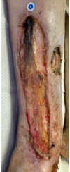 Patients A and B at initial visits — complex diabetic lower-extremity ulcers before treatment.
This case series was conducted as part of a hospital-based diabetic limb-salvage protocol in which three patients with diabetes and nine complex lower-extremity ulcers were treated with NPWTi-d and HOCl via ROCF-CC. The limb-salvage protocol also included glycemic control, offloading, revascularization when indicated, nutritional support, smoking cessation, systemic antibiotics, and sharp debridement.[3,5]
Following sharp surgical debridement, NPWTi-d was applied using a ROCF-CC dressing. Settings included instillation of hypochlorous acid every 2–3.5 hours, a dwell time of 10–20 minutes, continuous negative pressure at −125 mmHg, and dressing changes three times per week. These parameters are consistent with published NPWTi-d protocols using ROCF-CC in complex wounds.[3,6]
At each dressing change, the team used non-contact real-time fluorescence wound imaging to visualize regions of high bacterial load and non-contact near-infrared spectroscopy (NIRS) to measure perfusion/oxygenation (StO₂) in and around the wound.[7,8]
Results: Fast Conversion, Zero Amputations
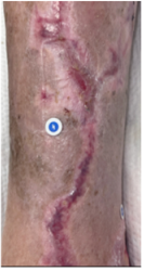 Patient B fully re-epithelialized 18 weeks after starting NPWTi-d therapy.
At baseline, wound volumes ranged from 4.6 to 49.2 cm³, with nonviable tissue covering between 15% and 100% of the wound surface.[3]
Under NPWTi-d with HOCl and ROCF-CC, all nine wounds converted to at least 90% coverage with clean granulation tissue in an average of 24.1 days. No patient required amputation during the observation period, and all limbs were salvaged.[3,4]
These findings align with other case series and retrospective analyses in which NPWTi-d with ROCF-CC has been associated with accelerated wound-bed preparation, earlier readiness for grafting, and limb preservation in high-risk diabetic and lower-extremity wounds.[4,5]
Mechanism: Hydromechanical Tissue Removal
NPWTi-d combines the mechanical benefits of negative pressure with cyclical instillation and dwell of a wound solution. During the instillation phase, the HOCl solution saturates the wound bed and helps loosen slough, fibrin and biofilm; during the dwell time, it interacts with devitalized tissue and bacteria; and during suction, the solution and solubilized debris are evacuated through the ROCF-CC's through-holes. This cycle promotes hydromechanical removal of nonviable tissue while maintaining an optimally moist wound environment.[6,9]
The case series emphasizes that this hydromechanical effect, delivered through the ROCF-CC contact layer, can decrease the need for repeated operative debridement and rapidly convert an infected, stalled wound into a clean, granulating base suitable for cellular, acellular or matrix grafting materials.[3,10]
Clinical Implications
For diabetic patients at high risk of limb loss, rapidly converting infected, necrotic ulcers into healing wounds is critical for avoiding major amputation. Integrating NPWTi-d with HOCl and ROCF-CC into a multidisciplinary limb-salvage protocol may reduce amputation risk by combining aggressive bioburden control, ongoing wound-bed preparation, and optimized perfusion.[3,5]
The use of fluorescence imaging to identify areas of high bacterial load and NIRS to assess tissue oxygenation provides actionable, point-of-care data to guide escalation or de-escalation of treatment. Clinical studies suggest that routine fluorescence imaging reduces unnecessary antimicrobial use while improving healing, and that NIRS-derived oxygenation metrics correlate with wound-healing trajectories in ischemic limbs.[7,8]
Workflow-wise, scheduled instillation every 2–3.5 hours automates wound cleansing between nurse visits, while dressing changes three times per week balance staff workload with close clinical monitoring.[4,6]
Visual and Practical Insights
A summarized table from the study lists each wound's initial size, percentage of nonviable tissue, and time to 90% granulation, illustrating how even wounds with extensive necrosis (up to 100% nonviable tissue) can be converted to healthy granulation within roughly one month when NPWTi-d is combined with systemic and local limb-salvage measures.[4,5]
Summary
This case series demonstrates that NPWTi-d with hypochlorous acid delivered via ROCF-CC can function as a form of automated hydromechanical debridement within a structured diabetic limb-salvage protocol. In nine high-risk diabetic lower-extremity ulcers, wounds transitioned from heavily colonized and necrotic to ≥90% clean granulation in an average of 24.1 days, with no amputations required. These early data, together with growing literature on NPWTi-d, fluorescence imaging, and NIRS, support considering this combined approach for select complex diabetic limb-threatening wounds.[3,6]
References
-
Armstrong DG, Boulton AJM, Bus SA. Diabetic foot ulcers: a review. N Engl J Med. 2023;389(2):e3. PubMed
-
McDermott K, Fang M, Boulton AJM, Selvin E, Hicks CW. Etiology, epidemiology, and disparities in the burden of diabetic foot ulcers. Diabetes Care. 2023;46(1):209–221. Diabetes Journals
-
Alonso MC, Singh J, Key D. Hydromechanical removal of nonviable tissue with negative pressure wound therapy and instillation to assist limb salvage. SAWC Spring 2025, Abstract CS-008. SAWC
-
McElroy EF, et al. Use of negative pressure wound therapy with instillation and dwell time using a reticulated open-cell foam dressing with through holes. Int Wound J. 2019;16(5):1188–1194. PubMed
-
Ioannidis O, et al. Tailored negative pressure wound therapy with instillation in patients with microangiopathy. World J Emerg Surg. 2025. BioMed Central
-
Kim PJ, Attinger CE, Steinberg JS, et al. Negative pressure wound therapy with instillation: international consensus guidelines update. Int Wound J. 2019;16(1):19–24. PMC
-
Armstrong DG, et al. Point-of-care fluorescence imaging reveals extent of bacterial load in diabetic foot ulcers. Int Wound J. 2023;20(2):554–566. PMC
-
Račytė A, et al. Oxygen saturation increase in ischemic wound tissues during therapy monitored by NIRS. Biomedicines. 2024;12(8):1805. MDPI
-
Téot L, Boissiere F, Fluieraru S. Novel foam dressing using NPWT with instillation to remove thick exudate. Int Wound J. 2017;14(5):842–848.
-
Price N, et al. Routine fluorescence imaging to detect wound bacteria reduces antibiotic use and antimicrobial dressing expenditure while improving healing rates. Diagnostics. 2020;10(11):927. MDPI